Few things are more frustrating than hearing that your MRI does not look bad enough to explain your pain. Patients often leave those conversations feeling dismissed or confused. Others have the opposite experience: the MRI report sounds alarming, yet their symptoms do not fully match what the scan seems to suggest.
At ATL Regen, we see this mismatch all the time. Imaging matters, but it does not tell the whole story. A scan is one piece of the puzzle. Pain is influenced not only by structure, but also by inflammation, tissue sensitivity, movement mechanics, nerve irritation, compensation patterns, and function.
What MRI is good at
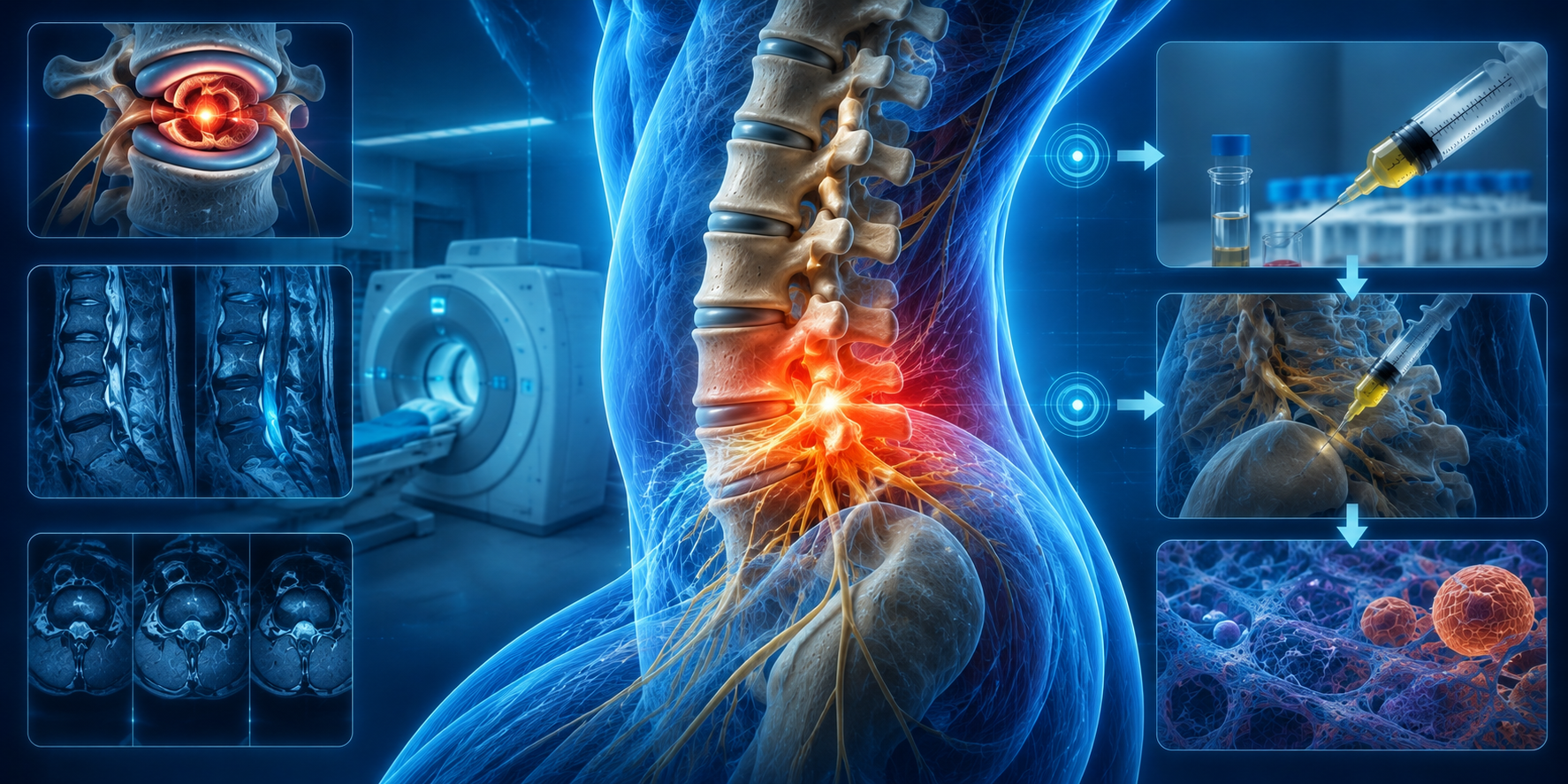
MRI is a useful tool because it can show disc bulges or herniations, degenerative changes, tendon or soft tissue injury, joint abnormalities, inflammation in some structures, and narrowing around nerves or the spinal canal. These are meaningful findings. MRI can help confirm whether there is a structural issue that fits the patient's symptoms.
What MRI cannot do on its own
MRI cannot fully show how irritated a tissue feels day to day, whether a visible abnormality is the active pain generator, how muscles are compensating, how the body moves under load, how chronic pain has affected sensitivity, or whether the patient's daily limitations match the scan. That is why a scan should support the clinical picture, not replace it.
Why symptoms and imaging do not always match
Many adults have degenerative changes, small tears, or disc bulges that cause little or no pain. A dramatic-looking scan may not represent the main pain generator. A relatively small structural issue can produce major symptoms if nerves or tissues are highly irritated. Muscle-based pain, guarded movement, and compensation can create significant pain with relatively mild imaging findings. The body's pain response involves the nervous system, which is not fully captured by a static structural image.
Why this mismatch is so important
When patients are told to trust only the scan, two problems can happen. Some patients with real pain are dismissed because their MRI looks mild. Others are pushed toward invasive treatment because the imaging looks dramatic, even when it may not fully explain the pain pattern.
What better correlation looks like
At ATL Regen, we ask: Where is the pain exactly? What movements provoke it? Is there radiating pain, numbness, or weakness? What does the physical exam show? Does the MRI finding line up with the symptoms and exam?
This process turns imaging into something more useful than a list of abnormalities.
Common scenarios patients experience
Patients often say their MRI is normal but they still hurt — in these cases pain may be coming from myofascial structures, inflammation, biomechanical overload, or another source not easily captured by the scan. Others say their MRI looks terrible but they are functioning okay — not every abnormality is equally symptomatic. And sometimes the most dramatic MRI finding is not the structure causing the patient's actual symptoms.
Imaging is most useful when it supports the story
MRI is extremely helpful when it is interpreted within the patient's real-world pattern. It can guide treatment when the level of nerve irritation makes sense clinically, the disc or joint abnormality matches the symptoms, the patient's exam findings support the image, and other causes have been considered. In that context, imaging can make treatment more precise.
Where PRP fits into the imaging conversation
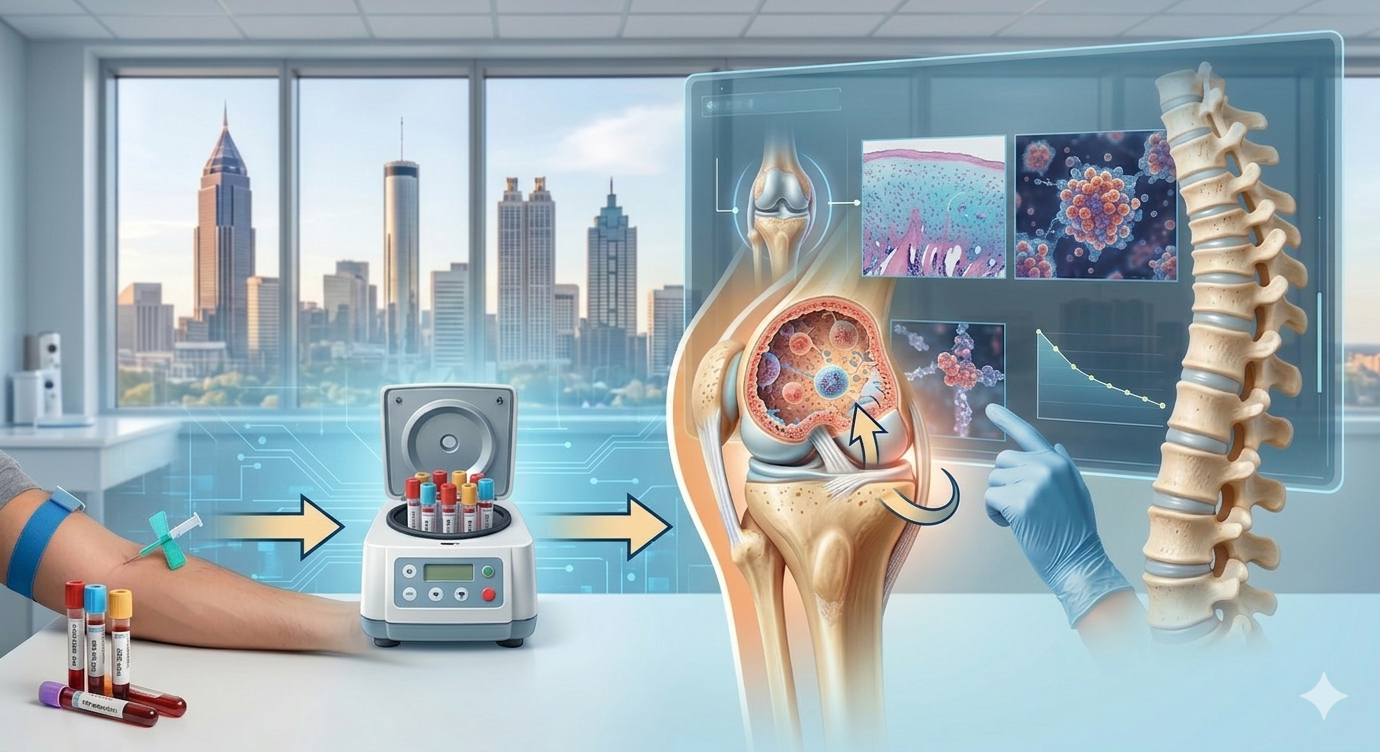
PRP is sometimes discussed when a scan shows a plausible tissue target and the patient wants to know whether a non-surgical option exists. But the presence of an MRI finding alone is not enough. PRP decisions still depend on diagnosis, function, symptom correlation, and treatment goals.
Schedule a consultation
If you have been told your MRI does not explain your pain, or if you are trying to understand whether your imaging truly matches your symptoms, ATL Regen can help put the pieces together more clearly.